What does an ultrasound show is one of the most common questions patients ask before their scan — and it deserves a clear, direct answer.
An ultrasound creates real-time images of the soft tissues, organs, and structures inside your body using high-frequency sound waves. No radiation. No needles. Just a safe, non-invasive look at what's happening inside.
Here's a quick overview of what an ultrasound can show:
| Body Area | What the Ultrasound Reveals |
|---|---|
| Abdomen | Liver, gallbladder, kidneys, pancreas, spleen |
| Pelvis | Uterus, ovaries, bladder, prostate |
| Thyroid | Nodules, growths, abnormal tissue |
| Breast | Cysts vs. solid masses |
| Heart & vessels | Blood flow, valve function, blockages |
| Musculoskeletal | Tendons, joints, muscles |
| Pregnancy | Fetal growth, heartbeat, position, anatomy |
It can also detect conditions like gallstones, kidney cysts, blood clots, tumors, and more — and even guide procedures like needle biopsies in real time.
What it can't show as clearly: air-filled areas (like the lungs) or structures behind bone (like the brain in adults). For those, a CT or MRI may be a better fit.
Most people associate ultrasound with pregnancy scans — and that's a big part of what it does. But it's actually one of the most versatile diagnostic tools in medicine, used every day across dozens of specialties.
Our team at AMI performs diagnostic ultrasounds using advanced equipment, read by our fellowship-trained radiologists, so you get accurate results you can trust.
To understand what does an ultrasound show, it helps to know how the technology creates those flickering gray-and-white images on the screen. Ultrasound, also known as sonography, doesn't use light or radiation. Instead, it relies on the same principles of echolocation used by bats, dolphins, and even submarine sonar.
The process centers around a handheld device called a transducer. When we place this probe against your skin, it sends out high-frequency sound waves that are far too high-pitched for human ears to hear. These waves travel through your skin and bounce off internal structures like organs, blood vessels, and tissues.
Denser tissues, like a gallstone or a solid tumor, reflect more sound waves and appear lighter gray or white on the screen. Softer tissues or fluid-filled areas, like a cyst or the bladder, allow more waves to pass through and appear darker. The transducer catches these "echoes" and sends them back to the computer, which translates the electrical signals into a real-time visual image called a sonogram.
One of the greatest benefits of this technology is that it provides a "live" look. Unlike a static X-ray, an ultrasound shows movement—the beating of a heart, the flow of blood through an artery, or a baby moving in the womb. Because it uses low-power sound waves, there is no ionizing radiation involved, making it a preferred choice for many diagnostic needs. You can learn more about the technical overview of ultrasound from the Mayo Clinic.
While many people think of ultrasounds exclusively for pregnancy, we use them to investigate symptoms throughout the entire body. Because the technology excels at visualizing soft tissues, it is often the first imaging test ordered when a doctor needs to see what is happening behind the scenes.
Ultrasound is highly sensitive to changes in soft tissue and blood flow, which makes it useful across a wide range of medical specialties — from cardiology to obstetrics to musculoskeletal medicine.
| Tissue Type | Appearance on Ultrasound | Examples |
|---|---|---|
| Fluid | Black / Very Dark | Bladder, cysts, amniotic fluid |
| Soft Tissue | Mid-range Grays | Liver, spleen, muscles |
| Dense Tissue | Light Gray / White | Gallstones, kidney stones, bone surfaces |
| Air/Gas | Distorted / Hazy | Bowel gas, lung tissue |
When we perform an abdominal scan, we are looking for the source of unexplained pain, swelling, or infection. We can see the liver to check for fatty liver disease or tumors, the gallbladder to find gallstones or inflammation, and the kidneys to look for stones or cysts. It’s also an essential tool for checking the spleen and pancreas.
The thyroid gland sits at the base of your neck and regulates your metabolism. An ultrasound can show the size of the gland and identify nodules. We use it to determine if a growth is a simple fluid-filled cyst or a solid mass that might require further testing.
We use MSK ultrasound to look at your "moving parts." This includes tendons, ligaments, muscles, and joints. It’s excellent for finding rotator cuff tears in the shoulder, Achilles tendon issues, or trapped nerves like those found in carpal tunnel syndrome.
When we use ultrasound to look at the heart, the test is called an echocardiogram. It shows us the heart's chambers, valves, and walls. We can see how well the heart muscle is pumping and if the valves are opening and closing properly.
For vascular health, we use Doppler ultrasound. This specialized technique allows us to see and measure the speed and direction of blood flow through your arteries and veins. It is a vital tool for:
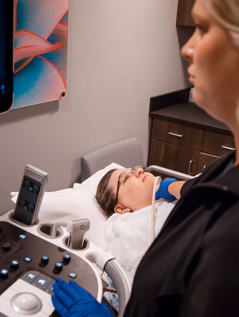
Not all ultrasounds are performed the same way. Depending on what your doctor needs to see, our team may use different methods to get the clearest possible image. At AMI Imaging, we offer a wide range of ultrasound services tailored to your specific health needs.
This is the most common type. We apply a cool, water-based gel to your skin and move the transducer over the area. It’s completely painless and used for most abdominal, pelvic, and pregnancy scans.
Sometimes, an external probe can't get close enough to the organs we need to see. In these cases, we use a specialized, thin probe that is inserted into a body opening:
While 2D ultrasound provides the flat, "slice-like" images used for most medical diagnoses, 3D ultrasound stitches those slices together to create a three-dimensional image. This is often used to look at fetal facial features or to evaluate uterine polyps. 4D ultrasound is simply a 3D image in motion, like a video.
Ultrasound isn't just for diagnosis; it’s also a guide. If you need a biopsy, our radiologists use ultrasound to see the needle in real-time, ensuring we collect tissue from the exact right spot. This makes procedures like breast biopsies or fluid drainage much safer and more accurate.
We know that medical appointments can be stressful, but an ultrasound is one of the most straightforward tests you’ll ever have. Most exams take between 30 and 60 minutes.
Your preparation depends entirely on what does an ultrasound show in your specific case.
We’ll have you lie comfortably on an exam table. We apply a clear gel to the area being scanned. This gel is vital because sound waves don't travel well through air; the gel creates a secure bond between your skin and the transducer. Our sonographer will move the probe back and forth, sometimes asking you to hold your breath for a few seconds to get a still image.
At AMI, we prioritize your comfort and convenience. We offer same-day appointments and provide quality imaging at outpatient prices, meaning you get the same high-level care without the "hospital markup."
Doctors choose imaging tests based on what they are trying to find. Ultrasound is versatile, but it has specific strengths and limitations compared to CT scans or MRIs.
| Feature | Ultrasound | CT Scan | MRI |
|---|---|---|---|
| Radiation | None | Low-dose X-ray | None (Magnetic) |
| Best For | Soft tissue, blood flow, pregnancy | Bone, lungs, complex trauma | Brain, spinal cord, ligaments |
| Cost | Most cost-effective | Moderate | Highest |
| Real-time? | Yes | No | No |
| Bone/Air | Limited | Excellent | Moderate |
Ultrasound is often the "first look" because it is safe, fast, and cost-effective. However, because sound waves cannot penetrate bone or travel through air, it isn't the right tool for looking at the brain inside the skull or the deep structures of the lungs. In those cases, our fellowship-trained radiologists might recommend a follow-up with a different modality. You can read more about ultrasound safety and research to understand why it is the preferred choice for many.
Yes. Diagnostic ultrasound has no known risks. Because it uses low-power sound waves instead of ionizing radiation, it is safe for pregnant women, infants, and people of all ages. However, we always recommend that ultrasounds be performed by trained medical professionals for a specific medical necessity to ensure the highest safety standards.
At AMI, we know you're anxious for answers. While the sonographer performs the scan, the images are interpreted by a board-certified radiologist. Your results are sent directly to your referring physician. You can also view your results and images through our secure online portal.
Ultrasound is a powerful tool in cancer detection, particularly for distinguishing between a harmless, fluid-filled cyst and a solid mass that could be a tumor. While it can't always tell if a mass is cancerous just by looking, it shows the mass's shape, location, and blood supply. We often use it to guide a biopsy needle to get a definitive answer.
When your doctor orders a scan, you have a choice in where you go. At AMI, we've built our reputation on providing the Lincoln community with sub-specialized, expert care in a warm and welcoming environment.
We are proud to be Lincoln's only ACR Designated Comprehensive Breast Imaging Center and the nation's only SphenoCath Certified Training Center. This means our radiologists aren't just generalists; they are fellowship-trained experts who specialize in specific areas of the body.
We believe that high-quality healthcare should be accessible. That’s why we offer outpatient pricing with no hospital markups and no surprise bills.
If you have a provider order and are ready to schedule, we invite you to request an appointment for an ultrasound today. Our team is ready to help you see what’s happening inside, with the care and expertise you deserve.
