Using ultrasound/MRI to track fatty liver disease gives doctors a safe, non-invasive way to see how much fat has built up in your liver and whether it's getting better or worse over time.
Here's a quick answer to what each test does:
Fatty liver disease, now more commonly called Metabolic dysfunction-Associated Steatotic Liver Disease, or MASLD, is a highly common liver condition in the United States, affecting up to 30% of adults. Most people have no symptoms at all. It's often discovered by accident during routine blood tests or an imaging scan ordered for something else entirely.
The good news is that modern imaging has come a long way. Our fellowship-trained radiologists use advanced ultrasound and MRI technology to detect liver fat early, monitor how your liver responds to lifestyle changes, and help your care team make confident decisions, all without a needle.

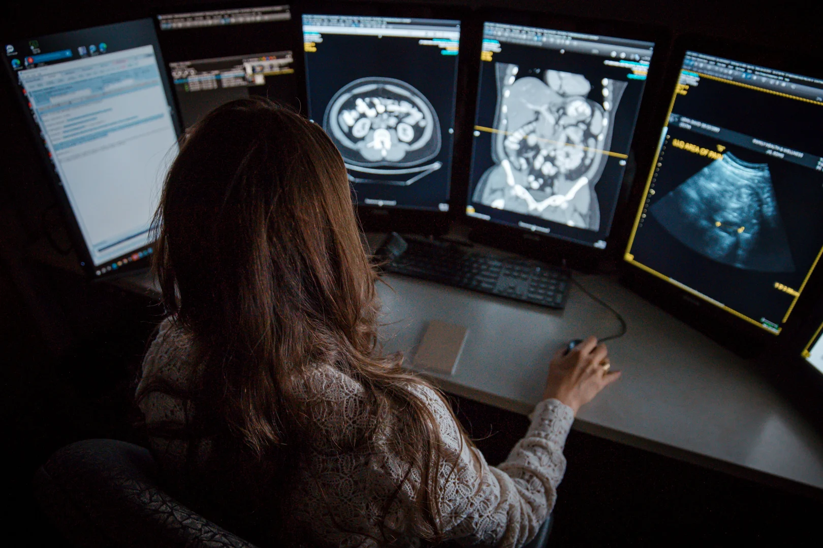
When evaluating diffuse hepatic steatosis (liver fat), choosing the right imaging modality is key. A landmark study published in the journal Clinical Imaging, Evaluation of diffuse liver steatosis by ultrasound, computed tomography, and magnetic resonance imaging, compared these three primary methods against liver biopsy results. The findings highlighted clear differences in diagnostic accuracy, safety, and reliability.
Standard ultrasound is highly valued as a safe, accessible first-line screening tool. However, it is fundamentally subjective and loses sensitivity when liver fat content is below 30%. It is also highly operator-dependent, meaning the results can vary depending on the person performing the scan.
Computed tomography (CT) can also identify moderate-to-severe liver fat, but it has notable drawbacks. CT scans are less accurate for detecting mild steatosis, and the measurements can be easily skewed by other liver conditions like excess iron or fibrosis. Furthermore, because a CT scan utilizes ionizing radiation, it is not recommended for routine, repeat monitoring or for pediatric patients. To learn more about how this technology works, you can read our overview on What is a CT Scan.
Magnetic resonance imaging (MRI), specifically opposed-phase MRI and quantitative techniques, consistently demonstrates an exceptionally high correlation with actual physical liver tissue samples. MRI can accurately detect even small amounts of liver fat, is highly reproducible, and completely avoids the radiation associated with CT.
A key aspect of managing MASLD or nonalcoholic fatty liver disease (NAFLD) is longitudinal monitoring. Because liver fat accumulation is highly responsive to lifestyle interventions, we need reliable tools to track your progress over time.
Research shows that losing just 3% to 5% of your body weight can significantly reduce liver fat, while a 7% to 10% weight loss can improve liver cell inflammation and even reverse early scarring. Non-invasive imaging allows us to see these positive changes happening in real time.
According to a study on Magnetic resonance imaging and spectroscopy for monitoring liver steatosis, standard clinical MRI protocols are exceptionally reliable for monitoring liver fat reduction. In patients undergoing a lifestyle-based weight loss program, MRI scans successfully documented a 50% or greater decrease in liver fat following modest weight loss. Because these scans can be repeated safely without radiation exposure, they are ideal for confirming that your treatment plan, diet, and exercise routine are successfully healing your liver.
For most patients, an abdominal Ultrasound is the first step when a primary doctor suspects a liver issue. It is widely available, fast, and entirely comfortable.
While conventional B-mode ultrasound has historically been limited by subjective visual grading, exciting technological advances have introduced quantitative ultrasound methods:
When your medical team needs a highly precise measurement of liver fat, quantitative magnetic resonance imaging is a gold standard. At Advanced Medical Imaging, we utilize advanced MRI Scans to provide highly detailed, objective data.
A leading tool for this is MRI-determined proton density fat fraction (MRI-PDFF). As highlighted in the clinical review Recent advances in noninvasive assessment of liver steatosis, MRI-PDFF has largely replaced complex magnetic resonance spectroscopy (MRS) in clinical practice because it is faster, commercially available, and covers the entire liver rather than a single small voxel.
Key clinical facts about MRI-PDFF:
Historically, a physical liver biopsy was a primary definitive way to diagnose and grade fatty liver disease. However, biopsies carry inherent limitations:
Thanks to modern imaging advances, we can often bypass the needle. As discussed in Goodbye biopsies, non-invasive diagnostics for fatty liver disease..., non-invasive diagnostics are rapidly becoming a preferred clinical standard. Major medical guidelines now recommend using advanced imaging like ultrasound/MRI to track fatty liver disease, reserving biopsies only for cases where the diagnosis remains unclear or when other complex liver disorders cannot be ruled out.
While tracking liver fat is important, a primary goal of managing MASLD is preventing the disease from progressing to liver inflammation (steatohepatitis) and permanent scarring (fibrosis). To protect Your Health, we use elastography to measure liver stiffness, which correlates directly with fibrosis.
There are two primary ways to perform elastography:
At Advanced Medical Imaging, we believe that tracking your liver health should be a seamless, reassuring experience. Our fellowship-trained radiologists specialize in sub-specialized abdominal imaging, ensuring your results are interpreted with an exceptional level of clinical expertise right here in Lincoln, Nebraska.
Whether you need a quick screening ultrasound or a highly precise quantitative MRI to monitor your liver's recovery, our team is dedicated to providing compassionate, high-quality care. We are conveniently located with same-day appointments available and are open 7 days a week to fit your schedule.
If you or your doctor would like to schedule a liver imaging study, please visit Advanced Medical Imaging or read more about our local Lincoln imaging services to schedule your appointment today.
