


A mammogram is a medical imaging exam used to look for changes within breast tissue. It can reveal signs of breast cancer before a lump or another physical symptom develops. Mammograms may also help a radiologist evaluate a new breast concern, an unusual clinical finding, or an area identified during an earlier test.
What is a mammogram? Understanding the purpose of the exam can make an upcoming appointment feel less uncertain. Patients often have questions about breast compression, radiation exposure, discomfort, results, and what happens if additional imaging is recommended.
Advanced Medical Imaging provides 3D digital mammography at its Women’s Center in Lincoln, Nebraska. AMI’s breast imaging services include routine screening, high-risk screening, diagnostic evaluation, and coordinated follow-up when further testing is needed. The practice combines advanced imaging technology with clear communication and personalized attention throughout the patient experience.
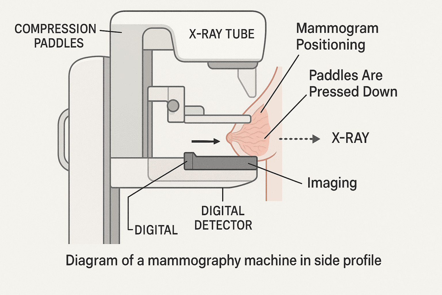
A mammogram is a low-dose X-ray picture of the breast. Mammography creates images that a radiologist reviews for masses, calcifications, tissue distortion, asymmetry, and other changes that may need closer evaluation. Because mammograms can detect some cancers when they're too small to feel, they remain the primary imaging test used for breast cancer screening.
During the exam, each breast is positioned on a flat imaging platform. A second plate lowers onto the breast and applies compression for a few seconds while an X-ray image is created. The technologist repeats the process from another angle and then performs the same steps on the other breast.
Compression spreads the breast tissue so structures are easier to see. It also helps reduce movement and allows the equipment to create a clearer image using a low radiation dose. The pressure may feel uncomfortable, particularly if the breasts are tender, but each image takes only a short time.
AMI uses 3D digital mammography, also known as digital breast tomosynthesis. During 3D mammography, the equipment obtains multiple low-dose images from different angles. Computer software then produces a series of thin breast images that the radiologist can examine in greater detail. AMI describes 3D digital mammography as its standard technology for annual screening at the Women’s Center.
Mammography does involve a small amount of radiation. The American College of Radiology reports that the radiation associated with one screening mammogram is comparable to roughly 26 days of natural background radiation. A medical provider can discuss any personal concerns, including pregnancy, previous radiation exposure, or the need for repeated diagnostic studies.
A mammogram and breast ultrasound serve different purposes. Mammography uses X-rays and is the main test for routine breast cancer screening. Ultrasound uses sound waves and may help determine whether a finding is solid or fluid-filled. It can also provide added information about a specific area. Ultrasound doesn't generally replace screening mammography, although it may be included in a diagnostic evaluation.
Breast MRI may be recommended for certain patients with a high risk of breast cancer or for selected diagnostic needs. The right test depends on symptoms, medical history, risk level, and the findings from previous imaging.
A routine screening appointment is designed for a patient who has no current breast symptoms and is due for regular breast cancer screening. The imaging itself often takes only several minutes, although check-in, changing clothes, positioning, and image review can make the full visit longer.
The technologist begins by confirming your medical information. You may be asked about previous breast surgery, biopsies, implants, hormone use, family history, and any recent changes. Accurate information helps the imaging team plan the exam and alerts the radiologist to factors that may affect interpretation.
You will undress from the waist up and receive a gown. The technologist positions one breast on the mammography unit and adjusts your body so the correct tissue is included in the image. You may be asked to move an arm, turn slightly, lift your chin, or hold your breath briefly.
For a typical screening mammogram, two standard views of each breast are obtained. Additional images may be necessary if positioning needs to be repeated, if the patient has implants, or if the technologist needs to include tissue that wasn't fully visible on the first image.
The technologist can't usually provide a diagnosis during the exam. Mammogram images must be interpreted by a radiologist, a physician trained to analyze medical imaging. The radiologist may compare the current study with prior mammograms to determine whether a finding is new, changing, or stable.
Comparison is particularly useful because breast tissue differs from person to person. A feature that appears unusual without prior images may have been present and unchanged for years. Patients who previously received mammograms elsewhere should ask whether those records need to be sent to AMI before the appointment.
Some people worry that the procedure will be painful. Experiences vary. Most patients feel pressure or temporary discomfort during compression rather than lasting pain. Breast sensitivity, implants, prior surgery, and individual anatomy may affect the experience. Tell the technologist if the pressure becomes difficult to tolerate. Minor positioning adjustments may help while still allowing the necessary images to be obtained.
A first mammogram can feel unfamiliar, but the process is structured and brief. Asking questions before imaging begins can help you understand what the technologist is doing and why each position is needed.
Screening and diagnostic mammograms use similar imaging technology, but they're ordered for different reasons. Knowing which appointment you need helps the imaging center schedule the appropriate amount of time and gather any required medical information.
| Type of Mammogram | Purpose | When It May Be Recommended | What to Expect |
|---|---|---|---|
| Screening mammogram | Looks for possible breast cancer before symptoms are present | Routine screening based on age and personal risk | Standard images of both breasts, followed by interpretation from a radiologist |
| Diagnostic mammogram | Examines a symptom, an unusual clinical finding, or an area seen on prior imaging | A lump, focal pain, nipple change, skin change, discharge, or abnormal screening result | Additional or magnified views focused on the area of concern, sometimes combined with ultrasound |
Screening mammography is preventive imaging. AMI states that women at average risk should begin screening at age 40, following the recommendation of the American College of Radiology for annual mammography beginning at that age. A patient’s doctor may recommend a different schedule based on family history, genetics, previous breast disease, chest radiation, or other risk factors.
A diagnostic mammogram is problem-focused. It may be ordered when a patient reports a new lump, localized breast pain, nipple discharge, nipple retraction, skin dimpling, swelling, or another change. It can also be used after a screening exam identifies an area that needs further assessment.
Diagnostic imaging may include extra views that aren't part of a routine screening study. These views can magnify an area, separate overlapping tissue, or show a finding from another angle. A breast ultrasound may be performed during the same visit when it can provide useful information.
A patient with a new symptom should tell the scheduling staff before booking a routine screening appointment. The center may need an order from a referring provider or may schedule a diagnostic visit instead. Choosing the correct appointment from the beginning can reduce delays and help the radiologist evaluate the concern properly.
Men can also undergo diagnostic mammography. Although male breast cancer is uncommon, a clinician may order mammography or ultrasound for a lump, nipple change, swelling, or another concerning symptom. Routine screening isn't commonly used for average-risk men, but individualized imaging may be appropriate for those with certain genetic or medical risks.
Preparation is simple, but a few steps can make the appointment smoother and help protect image quality.
Before your visit:
Don't skip an appointment because you have breast implants. Mammograms can still be performed, but the technologist needs to know about the implants in advance. Special positioning and additional images may be used to show as much breast tissue as possible.
Patients with limited shoulder movement, balance concerns, sensory sensitivity, or another condition that could affect positioning should also notify the center. Sharing this information beforehand gives the team an opportunity to plan appropriate assistance.
During your screening appointment, communicate openly with the technologist. Mention any location where you have felt a lump, pain, thickening, or another change, even if you're attending for routine imaging. A symptom may alter the type of study needed.
Patients sometimes assume they should cancel a mammogram when they have dense breast tissue. Density is determined from the images by the radiologist and is included in the mammogram report. Dense tissue is common, but it can make some findings harder to see because both dense tissue and many abnormalities appear white on an X-ray picture. Density may also affect breast cancer risk. Your doctor can help determine whether any additional imaging should be considered based on your full risk profile.
After the exam, a radiologist reviews the mammogram images and assigns an assessment. Many facilities use the Breast Imaging Reporting and Data System, commonly called BI-RADS, to organize findings and recommendations. The report is sent to the referring provider, and the patient receives a written summary of the results.
A normal mammogram means the radiologist didn't identify a suspicious change requiring additional evaluation. It doesn't mean breast cancer can never develop or that mammography detects every cancer. Continue following the screening schedule recommended by your medical provider and report new symptoms instead of waiting for the next routine appointment.
An abnormal mammogram doesn't automatically mean cancer. The term generally indicates that the radiologist needs additional information or has identified a finding that should be monitored or evaluated.
Possible next steps include:
Callbacks after screening are relatively common, especially after a first mammogram when no prior images are available for comparison. According to the American Cancer Society, fewer than 1 in 10 women called back for additional testing are found to have cancer. A callback often means the radiologist needs clearer images or wants to examine an area with another type of test.
Mammograms can show calcifications, masses, asymmetry, and architectural distortion. Some findings are clearly benign, while others require further assessment. Breast density is also documented because it affects how the images look and may influence discussions about risk.
Waiting for follow-up can cause understandable anxiety. Ask when results will be available, how they will be delivered, and whom to contact with questions. Complete recommended imaging promptly so the radiologist can obtain the information needed to make an assessment.
A mammogram provides information that can't be obtained through a physical exam alone. Whether you're preparing for your first routine screening, returning for annual mammography, or seeking evaluation of a new concern, receiving the correct type of imaging matters.
Advanced Medical Imaging’s Women’s Center offers 3D digital mammography, high-risk breast screening, and diagnostic breast imaging in Lincoln. AMI is an ACR-accredited Breast Center of Excellence and the first in Lincoln to be ACR accredited as a Comprehensive Breast Imaging Center. Same-day results are available in certain situations, subject to restrictions.
AMI’s radiologists specialize in breast screening and diagnosis, allowing care recommendations to reflect the patient’s risk level, symptoms, prior findings, and medical history. When additional testing is needed, the Women’s Center can help coordinate appropriate imaging and follow-up.
Women at average risk should speak with their provider about beginning annual mammograms at age 40. Patients with a family history of breast cancer, known genetic risk, previous breast disease, or prior chest radiation may need an earlier or different plan.
Contact Advanced Medical Imaging to request a mammogram in Lincoln, Nebraska, or to ask which type of breast imaging appointment is appropriate for your needs. A clear plan, experienced interpretation, and supportive care can help you approach breast screening with greater confidence.
.png)


