Dense breast tissue explained simply: your breasts are made up of two main types of tissue — fibroglandular tissue (the glands and connective fibers) and fatty tissue. When you have more fibroglandular tissue than fatty tissue, a radiologist will classify your breasts as dense on your mammogram report.
Here's what you need to know at a glance:
If your mammogram report mentioned dense breast tissue, you are not alone and you are not in danger simply because of that finding. But it is worth understanding — and worth discussing with your care team.
When you receive your mammogram report, you might see a specific section dedicated to breast density. At Advanced Medical Imaging, our fellowship-trained radiologists look at your X-ray images to determine the ratio of "white" tissue to "dark" tissue. On a standard mammogram, fatty tissue appears dark and transparent, while fibroglandular tissue appears solid white.
The challenge is that breast cancer also appears white on a mammogram. This creates what we call a "masking effect." Imagine trying to find a cotton ball in a bucket of milk—that is often what it feels like for a radiologist to look for a small tumor inside very dense tissue. Because of this, we use a standardized system to tell you and your doctor exactly how much dense tissue we see.
This system is called BI-RADS (Breast Imaging-Reporting and Data System), developed by the American College of Radiology. It ensures that whether you are seen in Lincoln or elsewhere, your results are communicated clearly. You can find more dense breasts: answers to commonly asked questions through the National Cancer Institute to help prepare for your next appointment.
Radiologists divide breast density into four distinct categories. Understanding which bucket you fall into helps us tailor your future screening plan.
If you are curious about how these categories affect your specific health journey, you can read more info about breast screening & diagnosis at our dedicated center.
There are two reasons why we pay so much attention to density. The first is the masking effect mentioned earlier—the "white on white" problem that can lead to a 40-50% miss rate in the most extreme cases. The second reason is more biological: breast cancer almost always originates in the glandular tissue, not the fat.
Because dense breasts have more of this glandular tissue, there is more "real estate" where cancer could potentially develop. This makes density an independent risk factor for breast cancer. In fact, women in Category D (extremely dense) have a 4 to 6 times greater risk of developing breast cancer than those with fatty breasts.
According to the CDC's guide on dense breasts, it is one of the strongest predictors of mammography's effectiveness. However, it is important to remember that having dense tissue does not mean you are more likely to die from breast cancer if it is caught early. It simply means we need to be more vigilant with our screening tools.
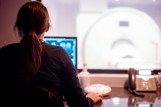
If you have dense breasts, a standard 2D mammogram may not be enough. At Advanced Medical Imaging, we utilize 3D mammography (also known as digital breast tomosynthesis) as a standard of care.
A 3D mammogram takes multiple images from different angles and reconstructs them into a "stack" of thin layers. This allows our radiologists to look through the dense tissue layer by layer, much like turning the pages of a book. This technology significantly reduces the "masking" effect and decreases the number of times we have to call patients back for additional images. You can find more info about 3D mammograms and why they have become the new standard for screening.
For women with high density or other risk factors, we often recommend supplemental screening. We're here for you every step of the way to help determine which of these tools is right for your unique biology.
Our team at the Women's Center can discuss these options with you during your visit.
Breast density is not a permanent setting; it can and does change throughout your life. Understanding these shifts can help you stay proactive.
In 2024, the FDA mandated that all mammography facilities in the U.S. notify patients about their breast density. This means you will receive a letter that clearly states whether your tissue is dense. When you see this, don't panic, it is an invitation to have a conversation.
At Advanced Medical Imaging, our fellowship-trained radiologists are sub-specialized in breast imaging. We are Lincoln's only ACR Designated Comprehensive Breast Imaging Center, which means we meet the highest national standards for safety and image quality. We provide quality imaging at outpatient prices, meaning you get world-class care without hospital markups or surprise bills.
We are open 7 days a week to fit your schedule, and we offer same-day appointments to reduce the time you spend waiting for answers. If your report shows dense tissue, we will work with you to perform a personal risk assessment and decide if supplemental screening is necessary.
Ready to take the next step in your breast health? Request an appointment at our Breast Center today.
Can I tell if I have dense breasts by how they feel?No. Density is a microscopic structural characteristic that can only be seen on a mammogram. Firm breasts are not necessarily dense, and soft breasts are not necessarily fatty.
Does breast density increase my risk of dying from cancer?Research shows that while density increases the risk of developing cancer and makes it harder to detect, it does not increase the risk of dying from the disease once it is diagnosed, provided you are receiving regular care.
Is a 3D mammogram enough if I have extremely dense breasts?While 3D mammograms are much better than 2D, women in Category D (extremely dense) should talk to their doctor about supplemental screening like ultrasound or MRI to ensure nothing is being missed.
How often should I get screened if I have dense breasts?For most women at average risk, we recommend an annual mammogram starting at age 40. If you have dense tissue, we simply might add an ultrasound or MRI to that yearly routine.
Does weight gain make my breasts less dense?Adding body fat can increase the amount of fatty tissue in the breast, which might change your BI-RADS category from C to B on paper, but it doesn't change the amount of glandular tissue you have or your underlying risk.
